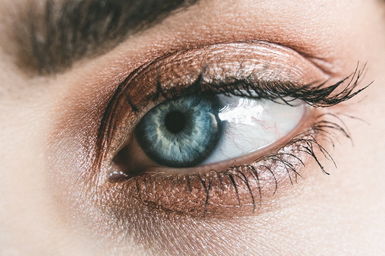
The Outside of Your Eyes
Eyelid
Description:
Your eyelid is a very thin fold of skin, adorned with a row of eyelashes. It is structurally supported by a robust tarsal plate, which gives your eyelids their definite shape. The eyelid also includes a layer of muscle responsible for the opening and closing of the eye.
Function:
The eyelids play a crucial role in protecting your eyes. The strong reflex action of your eyelids helps prevent foreign substances, such as dust and debris, from entering your eyes. Additionally, regular blinking spreads tears across the surface of your eyes, keeping them moist and ensuring clear, comfortable vision.
Sclera
Description:
The sclera is the white part of your eye.
Function:
The sclera is composed of tough tissue, which provides structural support and protection for your eyes. It acts as a sturdy shield, safeguarding the inner components of your eyes from damage.
Conjunctiva
Description:
The conjunctiva is a mucus membrane that covers the front part of your eyeball. It serves as a thin lining over the white part of your eye and receives oxygen supply from tiny blood vessels.
Function:
The conjunctiva acts as a protective seal, preventing objects like eyelashes and contact lenses from moving behind your eyeball. It also aids in spreading tears over the surface of your eyes, contributing to overall eye health.
Pupil
Description:
The pupil is a small black opening located in the centre of your iris.
Function:
The pupil allows light to enter your eyes and reach the retina. The muscles surrounding the pupil enable it to change size automatically in response to light levels. In darkness, the pupil enlarges to stimulate the peripheral retina, allowing better vision. Conversely, in bright light, the pupil contracts to minimise the intake of potentially harmful rays.
Iris
Description:
The iris is the coloured part of your eye, situated behind the cornea. It consists of tiny pigment cells known as melanin. The colour of your eyes is determined by the amount of melanin present in the iris. People with brown eyes have a higher concentration of pigment cells, whereas fair-skinned individuals with blue eyes have fewer pigment cells.
Function:
The iris contains a ring of muscular tissue that regulates the size of the pupil. It also acts as a barrier, separating the front and back parts of your eyes.
Cornea
Description:
The cornea is the transparent layer that covers the coloured part of your eye, including the iris, pupil, and anterior chamber (the space between the cornea and the iris). It lacks blood vessels, but it does have numerous nerves, making it a highly sensitive part of your eye.
Function:
The cornea plays multiple essential roles. One of its primary functions is to help focus incoming light onto the retina. Additionally, the high density of nerve fibres in the cornea helps detect foreign bodies, triggering a blinking reflex to remove any particles.
Tear Duct
Description:
The tear duct is located in the corner of your eye closest to your nose.
Function:
Any excess tears are drained from the surface of your eyes through the tear duct, also known as the puncta. This drainage system is why your nose runs when your eyes are watery from crying or allergies, and sometimes you can even taste the salt from your tears.
Lacrimal Gland
Description:
The lacrimal gland is situated under your upper eyelid.
Function:
Its primary function is to produce tears that cover the surface of your eyes, keeping them moist and clean.
The Inside of Your Eyes
Crystalline Lens
Description:
The crystalline lens is a transparent structure located behind your iris and pupil. It is held in place by numerous fibres, similar to strings attached to a puppet, which are in turn connected to a muscle.
Function:
The crystalline lens bends light onto the retina and works in tandem with the lens shape-changing ability, allowing you to focus at different distances. This process is known as accommodation.
Vitreous
Description:
The vitreous is a transparent gel that fills the chamber between your lens and retina.
Function:
Its main function is to maintain the shape of the eyeball and support the overall structure of your eyes.
Retina
Description:
The retina is a light-sensitive layer lining the inner part of your eyes. It consists of several layers and contains millions of light-sensitive receptors called rods and cones.
Function:
The receptors on the retina convert light into electrical signals that are transmitted via the optic nerve to the brain, where they are processed to create the images we see.
Macula
Description:
The macula is located in the centre of the retina at the back of your eyes. It contains the fovea, a small depression or pit at the macula’s centre, which has the highest concentration of photoreceptors in the retina.
Function:
The macula is responsible for detailed central vision, enabling tasks such as reading text, watching television, and recognising faces. The fovea provides the sharpest point of vision within the macula.
Optic Disc
Description:
The optic disc appears creamy-white and oval-shaped. It is the area where the optic nerve enters the eye and is located very close to the macula. The optic disc lacks photoreceptors, which results in a natural blind spot in each eye.
Function:
The optic disc is the starting point for all visual information received by the retina, which then travels to the optic nerve.
Optic Nerve
Description:
The optic nerve connects your eye to the brain. It functions like an electrical wire plugged into the brain.
Function:
Light detected by the retina is converted into electrical impulses, which travel along the nerve fibres from the optic disc to the brain. The brain then interprets these impulses to form the visual images we see.
Central Retinal Vein and Artery
Description:
The central retinal vein and artery are located at the back of your eyes along the retinal wall.
Function:
The retina requires a substantial amount of oxygen to function. The central retinal artery supplies blood to the retina, while the central retinal vein carries blood away from the retina, ensuring it receives the necessary nutrients and oxygen.