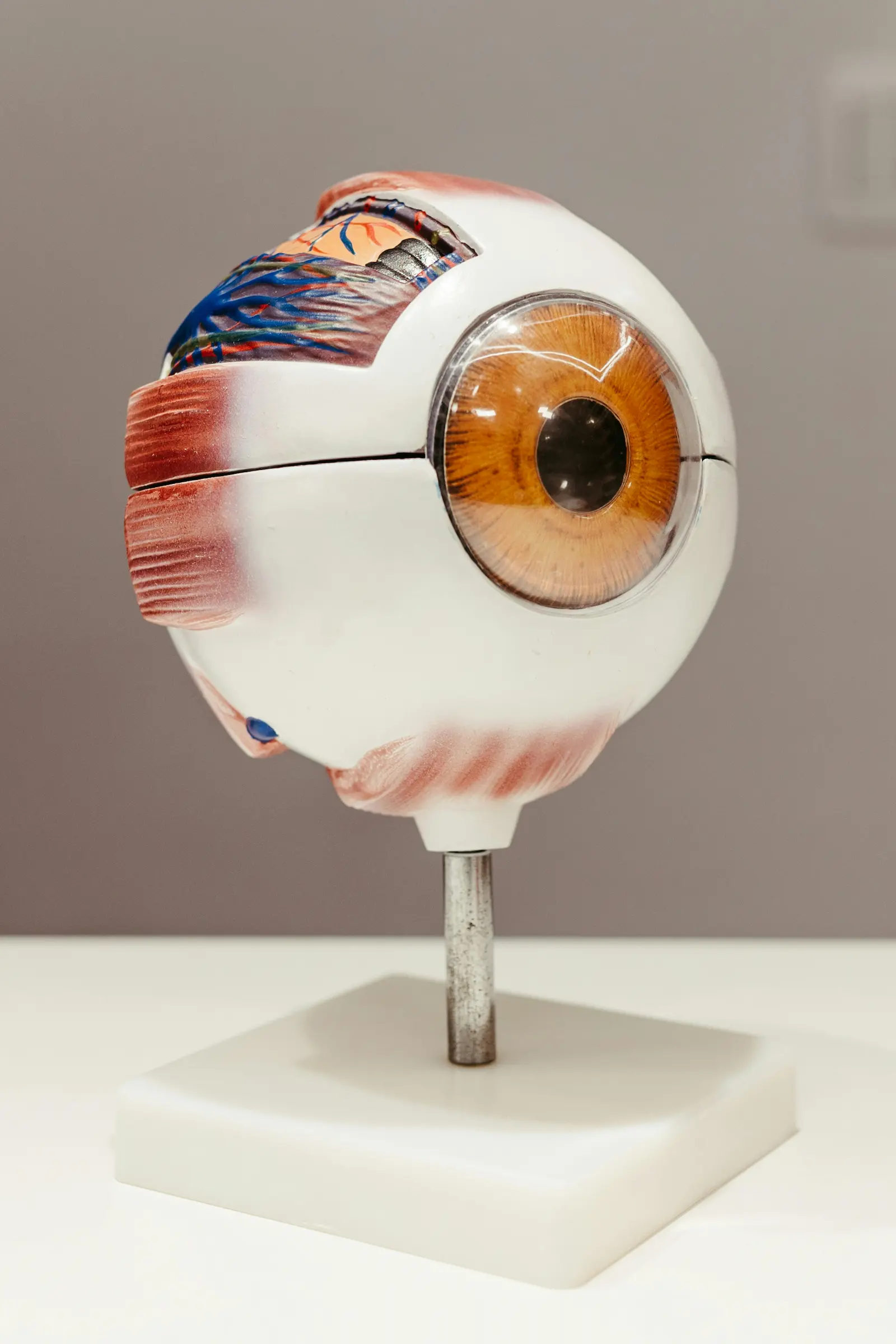
Introduction to Eye Anatomy
The human eye is a marvel of biological engineering, composed of various parts that work together to provide us with vision. Key components include the cornea, lens, retina, and the optic nerve. The cornea is the transparent, dome-shaped surface that covers the front of the eye and helps to focus incoming light. Behind the cornea is the lens, which further fine-tunes focus. The retina, located at the back of the eye, converts light into neural signals that the brain interprets as images. Understanding these parts is crucial for optometrists when examining patients.
Importance of Regular Eye Check-Ups
Regular eye check-ups are essential, not only for maintaining clear vision but also for the early detection of potential eye diseases such as glaucoma, cataracts, and macular degeneration. Regular visits to an optometrist can help prevent vision loss and other complications by catching problems early. An eye examination can also uncover other health issues, such as diabetes or high blood pressure, as these conditions often show early signs in the eyes.
Initial Patient Consultation
The first step in an eye exam involves a comprehensive consultation. The optometrist will ask about the patient’s medical history, current symptoms, and any family history of eye diseases. This initial consultation helps in tailoring the examination to the patient’s specific needs. Common issues such as blurred vision, headaches, and eye strain are discussed, providing essential insights into the patient’s condition.
Use of the Slit Lamp
One of the primary tools optometrists use is the slit lamp, a type of microscope with a bright light that allows a close-up view of the eye’s structures. The slit lamp helps in examining the cornea, lens, and anterior chamber for any signs of abnormalities such as cataracts or corneal damage. This tool is vital for diagnosing conditions that affect the surface and internal structures of the eye.
Retinal Examination Procedures
To examine the retina, optometrists often dilate the pupils using special eye drops. This dilation allows a comprehensive view of the retina and optic nerve. Using instruments like the ophthalmoscope and advanced imaging techniques, optometrists can detect conditions like retinal detachment, diabetic retinopathy, and age-related macular degeneration. These detailed examinations are crucial for maintaining overall eye health.
Farmilo’s Innovative Technology
Farmilo sets itself apart with innovative technology in eye care. Utilising state-of-the-art imaging equipment and diagnostic tools, Farmilo enhances the accuracy and efficiency of eye examinations. Their advanced technologies help optometrists to detect and treat eye conditions more effectively, ensuring patients receive the highest level of care. Farmilo’s commitment to innovation makes them a leader in comprehensive ocular health assessments.
In summary, examining the anatomy of the eye involves a series of detailed steps and cutting-edge tools. From initial consultations to sophisticated retinal imaging, each step ensures an accurate assessment of ocular health. Regular check-ups and the use of advanced technology like that offered by Farmilo can significantly improve vision care and overall eye health.